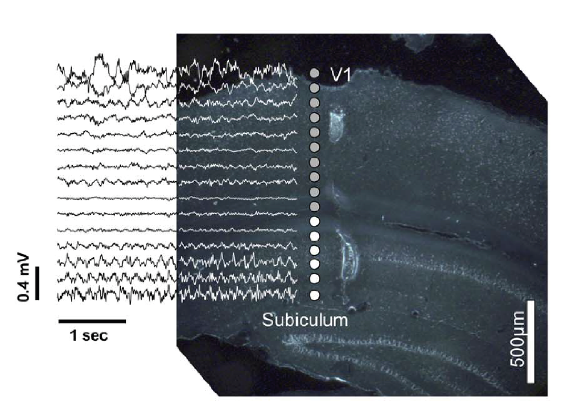
This project aims to develop a standardized mouse EEG approach, similar to a human standardized system, with defined electrode positions and methodology to facilitate comparisons between data from different studies and mouse strains. This aims for effective, comprehensive sensory phenotyping of transgenic mouse lines, especially for multisensory testing and a topographic overview of activity. As a stand alone, it allows to assess sensory capabilities before more complex experiments.
Topographical information is useful for testing auditory, visual or somatosensory cross-modal changes in the cortex, for example in deaf or blind mice strains, in order to study cross-modal plasticity. Further, this potentially allows to establish useful neural EEG markers for the use in mice, as a tool to better understand variability and effects of genetic and pharmacological influences. Recently, we demonstrated the feasibility for measuring visual evoked potentials with mouse EEG (Land et al. 2019).
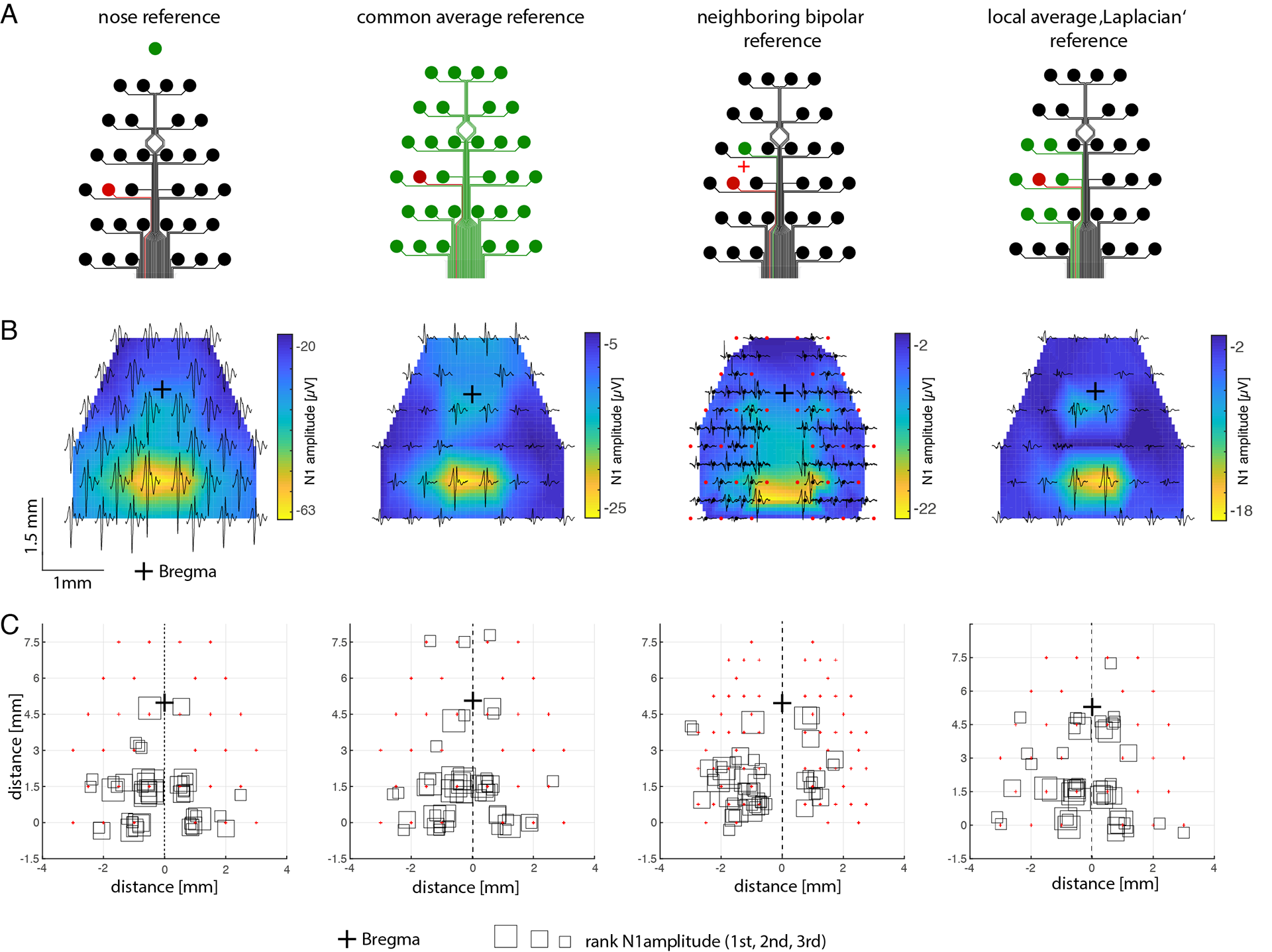
Mouse EEG with different configurations shows after re-referencing that this is usable for testing visual evoked potentials on different mouse strains (Land et al. 2019)
Auditory brainstem Response - The P-wave
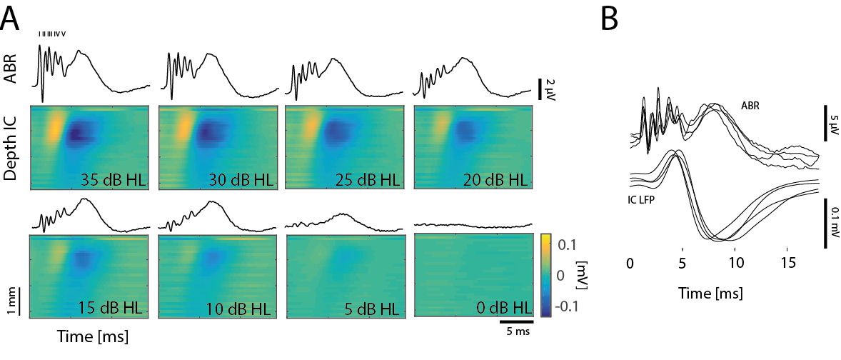
Mouse EEG can be used to assess biomarkers of different conditions, specifically often used to assess hearing status by measuring ABR waves. This provides an objective measure for the integrity of the auditory pathway along in the auditory brainstem. However, it is necessary to know the origin of the ABR waves, and this origin is still not perfectly clear for the later waves. One important structure is the inferior colliclus as a most important relay station of all auditory information to the cortex. One can identify a specific marker for IC activity in the ABR response when filtering the signal adequately. This slow component provides a non-invaisve measure of IC activity in the mouse, and correlates well with activity recorded within the IC (Land et al. 2016).
By combining invasive recrodings with surface recordings different components can be established in more detail, especially the contribution of the IC to the ABR activity in the mouse. This can then be used to phenotype more effciently, when testing auditory function in transgenic mouse strains.
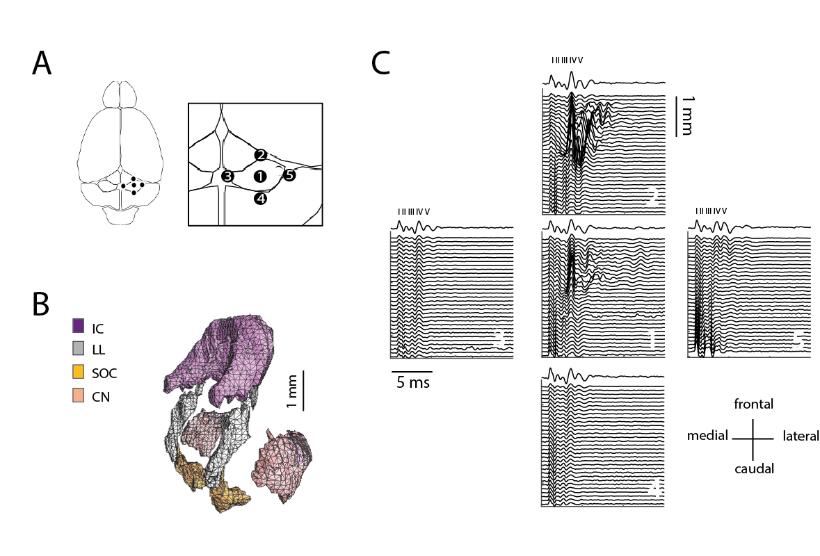
Mouse ABR and measured responses within the mouse inferior colliculus with a 32-channel multielectrode array. Latency help to determine possible generators for the ABR waves (Land et al. 2016)
.
References
Land, R., Kapche, A., Ebbers, L. & Kral, A. 32-channel mouse EEG: Visual evoked potentials. J. Neurosci. Methods 108316 (2019).
Land, R., Burghard, A. & Kral, A. The contribution of inferior colliculus activity to the auditory brainstem response (ABR) in mice. Hear. Res. 341, 109–118 (2016).
BRAIN STATES AND ANESTHESIA
General anesthesia is an interesting brain state. Similar to sleep, it is a reversible state, during which consciousness is switched off. However the brain is still active, and most notabely sensory information still travels through the sensory pathway and reaches the sensory cortex. However, we do not have awareness of this sensory information during general anesthesia.
Also similar to sleep, during anesthesia it is possible to observe different brain states, depending on the depth of anesthesia and the the type of anesthetic. Interestingly, allthough a large number of sensory neurophysiology experiments are performed during general anesthesia, it is intriguing how rarely processing during different anesthesia states is specifically addressed in studies.
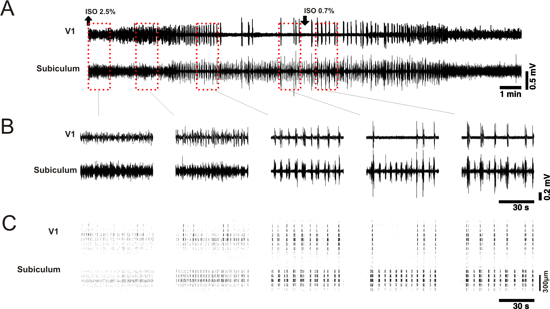
We are interested in sensory responses during different varying anesthetic depths. This is interesting for better control of measurements during general anesthesia of sensory evoked responses (Land et al. 2013). On the other hand, it allows to study effects of general anesthetics on the cortical network, and how it influences the dynamics in a level-dependent manner. This also may allow to understand better the mechanisms that are apparently suppressed during anesthesia, that are necessary for conscious experience. We have studied auditory and visual responses during isoflurane anesthesia in the visual cortex of mice (Land et al. 2012). We found non-linear effects during high anesthesia levels with isoflurane, where auditory stimulation evoked activity within the visual cortex.
References
Land, R., Engler, G., Kral, A. & Engel, A. K. Auditory Evoked Bursts in Mouse Visual Cortex during Isoflurane Anesthesia. PLoS One 7, e49855 (2012).
Land, R., Engler, G., Kral, A. & Engel, A. K. Response properties of local field potentials and multiunit activity in the mouse visual cortex. Neuroscience 254, 141–51 (2013).